The liver is a vital organ responsible for metabolism and detoxification. Understanding the differences between a normal liver, fatty liver disease, and alcoholic liver disease is essential for medical students and educators. By observing changes in liver color, size, shape, and the distribution of blood vessels and bile ducts, combined with the use of liver models, learners can better grasp the characteristics of liver diseases and improve clinical diagnostic skills.
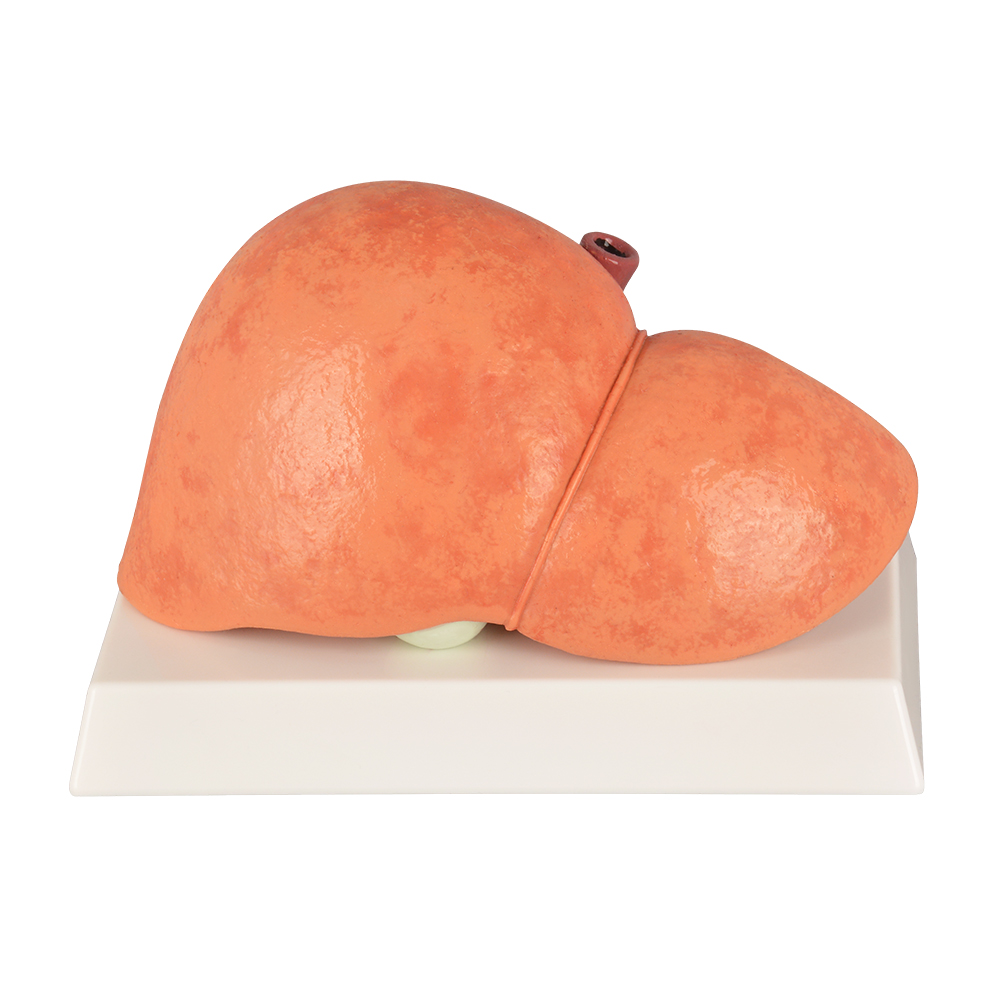
- Color: The normal liver has a reddish-brown color with a glossy surface, indicating good blood supply.
- Size: The adult liver is moderate in size, weighing approximately 1200–1500 grams, with sharp and clear edges.
- Morphology and Surface: The liver surface is smooth and regular, without nodules.
- Anatomical Segmentation: Liver lobes and segments are clearly defined for easy identification.
- Vascular and Biliary Distribution: The portal vein, hepatic artery, and bile ducts are regularly distributed with clear structures.
- Distinctive Features: Uniform liver texture and normal function.
- Surface Details: No fibrosis or nodules.
- Pathological Changes: None.
Fatty Liver (Non-Alcoholic Fatty Liver Disease, NAFLD)
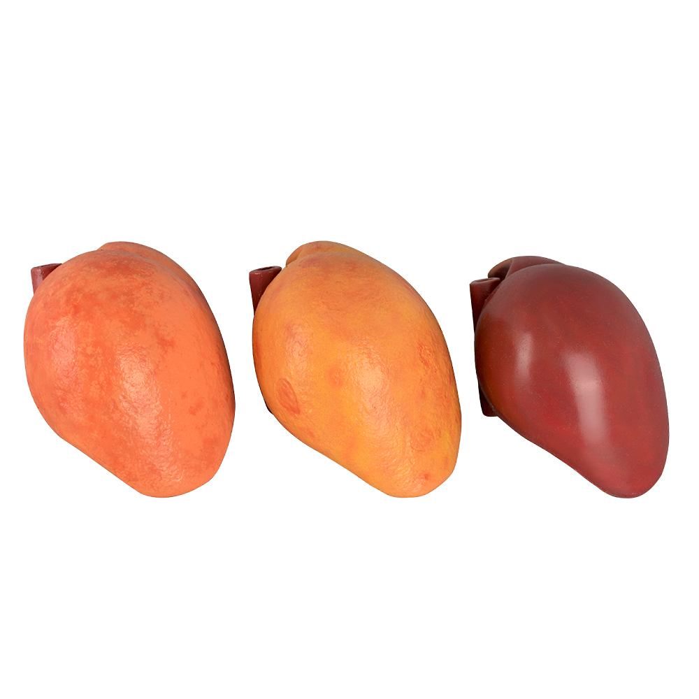
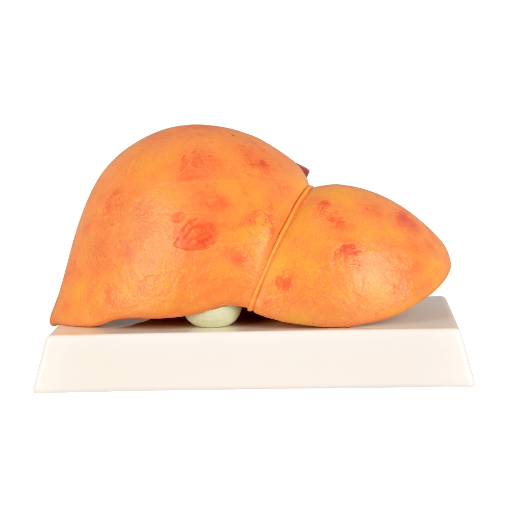
- Color: Fatty liver appears lighter, pale yellow or greasy yellow, due to fat accumulation inside liver cells.
- Size: The liver enlarges and feels softer on palpation.
- Morphology and Surface: The surface remains mostly smooth, but the edges become more rounded and blunt.
- Anatomical Segmentation: Liver lobes are mostly intact but may appear slightly blurred due to fat infiltration.
- Vascular and Biliary Distribution: Large vessels maintain normal structure, though fat deposits may compress smaller vessels.
- Distinctive Features: Fat accumulation causes liver enlargement and lighter color.
- Surface Details: Smooth surface without nodules.
- Pathological Changes: Fat droplets accumulate in hepatocytes with minimal or no inflammation.
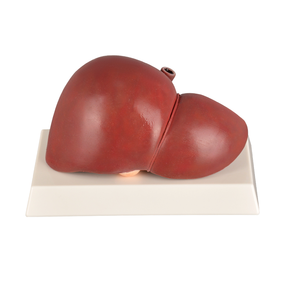
- Color: The liver color darkens to a deep red or brownish tone, often uneven due to inflammation and tissue damage.
- Size: Early stages show liver enlargement, but in advanced disease (cirrhosis), the liver shrinks and becomes firm.
- Morphology and Surface: The surface becomes coarse and irregular, with nodules and fibrous plaques; edges thicken and blunt.
- Anatomical Segmentation: Liver lobes and segments may become indistinct or lost due to fibrosis.
- Vascular and Biliary Distribution: Fibrosis distorts and compresses vessels and bile ducts, often leading to portal hypertension.
- Distinctive Features: Presence of fibrous nodules and a hardened liver texture.
- Surface Details: Numerous irregular nodules connected by fibrous tissue.
- Pathological Changes: Hepatocyte degeneration, necrosis, inflammation, and fibrosis
Fatty Liver: Alcoholic vs. Non-Alcoholic
| Aspect |
Non-Alcoholic Fatty Liver Disease (NAFLD) |
Alcoholic Fatty Liver Disease (AFLD) |
| Cause |
Related to metabolic syndrome, not caused by alcohol |
Caused by long-term excessive alcohol consumption |
| Clinical Presentation |
Mostly asymptomatic or mild discomfort |
Fatigue, right upper abdominal pain, significant liver function abnormalities |
| Key Diagnostic Factors |
No significant alcohol history; presence of metabolic abnormalities |
Positive history of alcohol use; obvious liver function impairment |
| Treatment |
Lifestyle changes (weight loss, exercise) |
Alcohol cessation and nutritional support |
Clinical Presentation Summary
Generally asymptomatic, with normal liver function tests and no abnormalities on physical examination.
Most patients are asymptomatic but may experience mild fatigue or right upper abdominal discomfort. Liver function tests are usually normal or mildly elevated. Often detected during routine health exams or imaging such as ultrasound.
Patients may present with fatigue, loss of appetite, nausea, right upper quadrant pain, and jaundice. Laboratory tests often show abnormal liver function with elevated serum transaminases (AST typically higher than ALT). Advanced cases may show signs of liver cirrhosis and liver failure.
Conclusion
Recognizing differences in liver color, size, morphology, and vascular structures, along with clinical symptoms, enables better differentiation between normal liver, fatty liver, and alcoholic liver disease. The use of anatomical and pathological liver models enhances learning by providing hands-on visualization of disease changes, which is invaluable for medical education and clinical training.


